What You Need to Know About Dental X-Rays
December 29, 2019
Dental x-rays are now an essential part of modern dentistry. They provide us with a huge amount of diagnostic information. They allow us to see what is going on under the gum, inside the tooth and in the surrounding bone. X-rays are very safe.
An x-ray works in much the same way as a camera. The difference is that it uses a small dose of radiation instead of light. This shines through your jaw and records an image on a film which we can then view. The radiation dose with conventional films is very small. It has been compared to the same quantity of radiation you receive while flying on an international flight. Using the latest digital equipment we can further reduce your radiation exposure to 10% of conventional x-rays.
The advantages of digital x-rays include:
- Reduced radiation exposure to 10% of conventional x-rays.
- X-rays can be viewed on computer immediately.
- Better for the environment as no chemicals are needed to develop films.
- Safer for staff as they are not exposed to poisonous chemicals.
- Images are in digital format so we easily send them to specialists or to patients who require copies.
Intraoral x-rays
These x-rays are placed inside your mouth and show us detailed images of the tooth and its surrounding structures. They very helpful for diagnosing the following:
Tooth decay
Images show if decay is present, where it is and how deep it is. By comparing successive x-rays we can also see how fast decay is progressing. Sometimes it is not advancing at all and if the surface of the tooth is still intact, we can watch and repair it with careful home care and fluoride application. Routine ‘bite-wing’ x-rays of your back teeth should be taken every 2 years for a healthy individual, more frequently if your teeth are assessed as being at increased risk of decay.
Problems with existing fillings
X-rays allow us to see in between teeth and under existing dental work. We can see filling overhangs, poor fitting crowns and bridges and leaking or broken fillings that may need repair or replacement.
Gum disease
We can accurately see bone levels around each tooth and where bone has been lost due to gum disease. Again we can compare successive x-rays to tailor treatment and make sure the disease is under control. In some people the calculus (hardened plaque) is so thick on their teeth you can see it on an x-ray.
Root canal treatment
After 10 days of dental infection, early abscesses can become visible in the bone as a shadow on x-ray. Well established dental infections are usually more obvious. These indicate the nerve of a tooth is dying and root canal treatment is needed. During root canal treatment x-rays are used to measure the length of the root and make sure all the infected nerve is removed. Finally, review x-rays are taken after completion, to make sure the site is healing and treatment is a success.
Accidents
X-rays are a vital part of assessing teeth after accidents. We look for cracks and fractures, teeth knocked out of position, damage to adult teeth below and infections. They are also used in future reviews to make sure growth is normal and injuries are healing correctly.
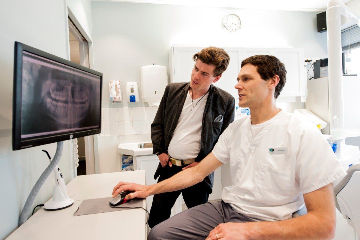
Panoramic x-rays
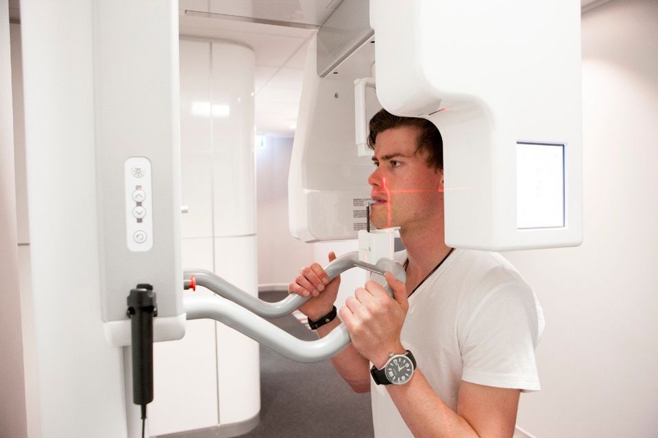
The jaw is a curved shape, similar to a horse shoe. A panoramic x-ray provides a flat, two dimensional image of this half circle. The film does not go inside your mouth but sits inside the machine instead. To minimise radiation exposure and provide an instant computer image, our panoramic x-ray machine is also completely digital.
It captures all the upper and lower jaw and provides information on:
- impacted teeth (teeth which haven’t come through into the mouth)
- extra and missing teeth
- bone loss from gum disease
- accident assessment including broken jaws
- dental implant assessment
- denture assessment
- orthodontic assessment
- the jaw joint (TMJ)
- the sinuses and nasal cavities
- cysts
- cancers and tumours
- calcifications in the carotid arteries (indicating atherosclerosis and increased risk of heart attack)
- diagnosis of dental abnormalities
Panoramic x-rays are excellent at showing us the ‘big picture.’ However intra-oral x-rays are still needed to see fine detail such as tooth decay. Panoramic x-rays are routinely taken as part of our ‘New Patient Examination’ and in other cases when indicated.
Safety: lead shields and pregnancy
When using the older x-ray machines small amounts of the main x-ray beam used to bounce off and ‘scatter’ sideways, potentially exposing the other parts of the body. In the past some dentists used lead shields to protect you from this radiation. Our modern equipment uses filters, collimators and a decreased exposure time, so scattered radiation is almost completely eliminated. There is also a theory that lead neck shields can bounce the scatter radiation back to your body. For these reasons we do not routinely use them.
As a precaution, to give unborn babies the best protection, we do avoid x-rays for pregnant mothers unless absolutely necessary. The early embryo is such a small group of developing of cells we do not want to risk any radiation exposure at all. We do have a full body lead shield to protect in utero babies if needed. It is important to tell us if you think you might be pregnant before we take any x-rays.

There are situations where pain relief is needed, such as travelling in to see us with a sore tooth, or healing an infected or damaged tooth. We give you some guidelines on how best to manage dental pain.
A typical dental hygienist appointment will involve ‘debridement and prophylaxis’, the removal of a plaque and calculus from the surfaces of your teeth. We explain what may happen after this procedure.

We explain what to expect after a Root Canal treatment - how to optimise the success of the treatment and avoid possible complications.